Endoanal Ultrasound

Endoanal ultrasound (or transrectal ultrasound) utilizes the same imaging method as classic ultrasound - an elongated, rotating head sends ultrasound waves that reflect from the tissues that form and surround the anus, creating an image on the computer and allowing an assessment of the structure of all surrounding structures.
What is endoanal ultrasound?

What is endoanal ultrasound?

Transrectal ultrasound is the basic proctological examination performed when the doctor suspects changes in the anus and its surroundings, the full picture of which is impossible to assess using anoscopy.

One of the basic applications of endoanal ultrasound is to determine the course of perianal fistulas or the location of perianal abscesses, which may occur, among others, in patients with Crohn's disease.

It further enables the determination of damage to the anal sphincter muscles in cases of fecal incontinence in women after natural labor, the elderly, and after surgery around the anus.

Based on the transrectal ultrasound image, the stage of anal cancer is determined for the purpose of planning surgical treatment. Knowledge of the degree of tissue involvement by the tumor and selecting an appropriately conservative surgical method saves many patients the discomfort associated with the need to create an intestinal fistula (the so-called stoma).
Book a Visit

How is the examination done?

In order to perform the examination, the patient undresses from the waist down and then lies on the couch on the left side with the knees pulled up to the stomach. Then, in order to minimize the discomfort associated with the examination, the doctor inserts an ultrasound probe into the rectum, secured with an appropriate shield, and covered with gel.

The examination is not painful, but some patients may find it uncomfortable. If you experience severe pain during a transrectal ultrasound, you should inform your doctor who will stop the examination immediately.

The examination lasts 5-10 minutes, depending on the complexity of the changes observed. After the examination, the doctor can view the images from the examination in a 3D view, which allows the detection of even the smallest changes in the anus and its surrounding tissues.

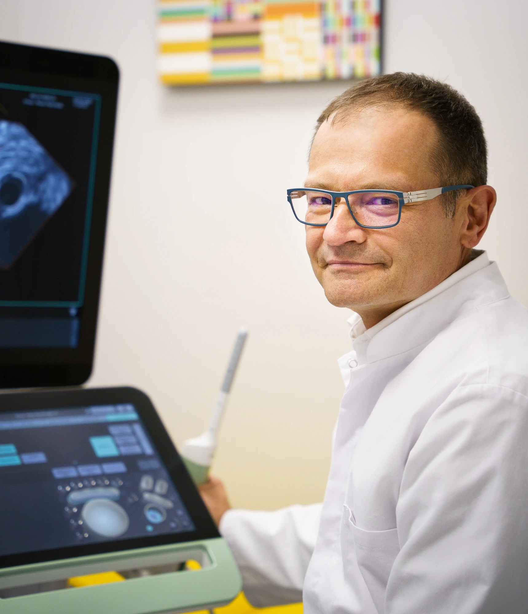
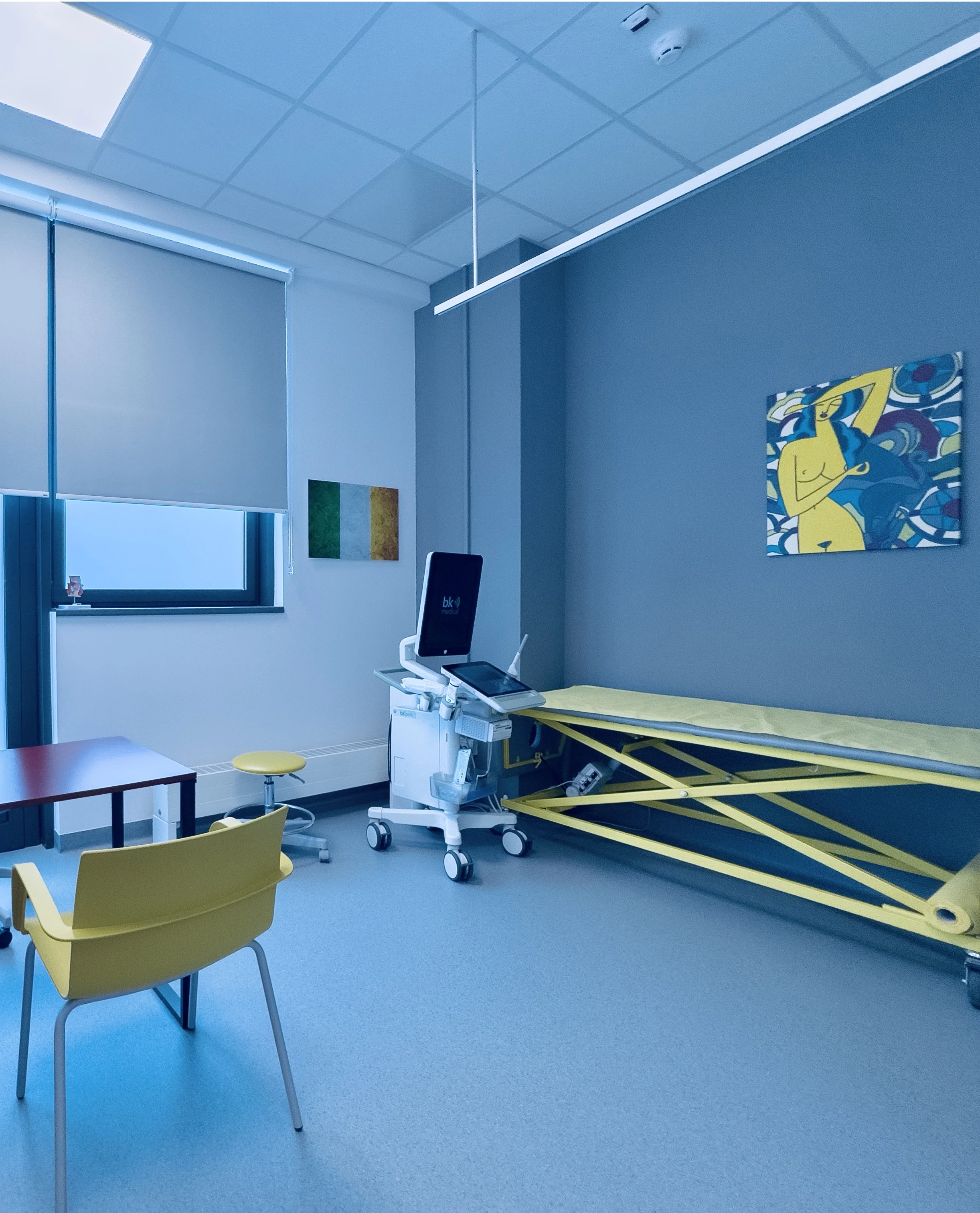
An advanced ultrasound scanner by BK Medical is used for examinations at the Melita Medical Center.

Book a Visit
How is the examination done?
When an endoanal ultrasound should be performed?

When an endoanal ultrasound should be performed?

The examination is usually recommended after consultation with a gastroenterologist, proctologist, surgeon, or oncologist who sees the need to assess the structures of the anal area in order to plan further treatment.

The indications for transrectal ultrasound are:

  • The presence or suspected presence of the perianal fistula or perianal abscess
  • Fecal and gas incontinence (after childbirth, anal surgery)
  • Presence of operable rectal cancer (for the determination of the surgery type)

How do get prepared for the examination?

You do not need to prepare in any special way for this examination. It may only be beneficial to refrain from eating a meal for about 2 hours before the visit, defecate before the examination (if needed) and take care of the hygiene of the anal area.

Book a Visit

Price list

Placement of a draining seton for fistula under intravenous sedation (analgosedation)
from 2000 PLN
Operation of low fistulas in analgosedation and local anesthesia
from 2500 PLN
3D ultrasound of the rectum to determine the course of the fistula tract
750 PLN
Book a Visit